Image analysis and multiparametric quantitative fluorescent microscopy reveal tissue changes between healthy and diseased human liver
- Principal Investigators:
- Prof. Marino Zerial
- Project Manager:
- Dr. Yannis Kalaidzidis
- HPC Platform used:
- NHR@TUD Taurus and Barnard
- Project ID:
- ht-10
- Date published:
- Researchers:
- Carlotta Mayer, Sophie Nehring, Michael Kücken, Hernán Morales-Navarrete, Mario Brosch, Meritxell Huch, Yannis Kalaidzidis, Lutz Brusch, Jochen Hampe, Marino Zerial
- Introduction:
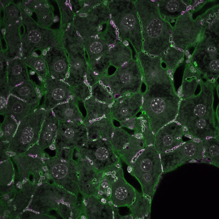
- The liver produces bile, which the intestine uses for digestion. For the transport of bile, the liver relies on a network of microscopic tubings, known as bile canaliculi, formed by liver cells called hepatocytes. When the outflow of bile to the intestine is blocked, it collects in the liver and can lead to serious liver disease. Researchers at the Max Planck Institute of Molecular Cell Biology and Genetics (MPI-CBG) in Dresden together with experts from the Carl Gustav Carus University Hospital (UKD) and the Department for Information Services and High Performance Computing (ZIH) at TU Dresden as well as further clinics in Germany and Oslo University Hospital in Norway found that high pressure in the bile canaliculi alters the structure of the liver tissue. Using deep image analysis, they found that elevated pressure leads to the obviation of apical bulkheads, structures known to reinforce the canaliculi. Subsequently, bile canaliculi enlarge into liver cell rosettes that are observed in many liver diseases. This study identifies pressure to be a potential common cause of various liver diseases with biliary obstruction. In the next step, we are applying the gained knowledge about bile pressure to better understand the development of liver cancer, again using HPC-scale image analysis of human liver samples.
- Body:
-
The research groups led by Marino Zerial, director at the MPI-CBG, and Jochen Hampe at the UKD, in collaboration with colleagues at the CIDS-Department for Information Services and High Performance Computing (ZIH) of TU Dresden and the Oslo University Hospital, have found initial evidence that bile stasis leads to excess pressure in the microscopic bile canaliculi of the human liver. Long-term overpressure could damage liver tissue and, consequently, lead to liver diseases such as primary sclerosing cholangitis (PSC) and primary biliary cholangitis (PBC).
Zooming into the bile cancaliculi using high-resolution microscopy, scientists now understand better what happens in the microcirculatory system of the liver tissue when it's under high pressure. Hepatocytes form transversal connections called “apical bulkheads,” stabilizing the bile canaliculi tubules. But when there's too much pressure from bile, the stabilizing supporting hepatocytes start to break down. This leads to the widening of the tubes and the formation of new structures called “rosettes.”
For decades, physicians have used liver rosettes as a marker in the diagnosis of cholestatic liver disease to detect these disorders. In these structures, abnormal liver cells arrange like rose petals around a small opening. Such rose-like structure can be identified in tissue samples. Why these structures form, however, was unclear up to today.
Thanks to state-of-the-art fluorescence microscopy techniques, the researchers understood that liver cell rosettes are cross-sections of the bile canaliculi suffering excess pressure due to bile stasis. The scientists have thus solved a mystery about the causes of the formation of these structures, which are considered a distinguishing feature of biliary congestion in liver tissue. This suggests a direct link between increased pressure in the bile canaliculi and cholestatic liver disease and improves our understanding of these diseases.
Carlotta Mayer, the first author of the study, explains, “We see liver cell rosettes already in the early stages of PSC in the tissue. Thus, they are potential diagnostic markers of early stages of PSC and other liver diseases.” The research findings suggest that excess pressure in the bile canaliculi may also play a role in other liver diseases, such as hepatitis. This could open new avenues for research into liver diseases.
“This work is an excellent example of the need for interdisciplinary research bridging medicine and cell biology. We now need to focus on the molecular mechanism of liver rosette formation to open new perspectives for treatment of these diseases,” emphasizes the head of the research group, Prof. Marino Zerial.
This study has been published in:
C. Mayer, S. Nehring, M. Kücken, U. Repnik, S. Seifert, A. Sljukic, J. Delpierre, H. Morales-Navarrete, S. Hinz, M. Brosch, B. Chung, T. Karlsen, M. Huch, Y. Kalaidzidis, L. Brusch, J. Hampe, C. Schafmayer, M. Zerial. Apical bulkheads accumulate as adaptive response to impaired bile flow in liver disease. EMBO Reports 24 e57181, 2023. https://doi.org/10.15252/embr.202357181 - Institute / Institutes:
- Technische Universität Dresden, Max Planck Institute of Molecular Cell Biology and Genetics (MPI-CBG)
- Affiliation:
- Technische Universität Dresden, Max Planck Institute of Molecular Cell Biology and Genetics (MPI-CBG)
- Image:
-